-

Harmona Abonales posted a new activity comment 1 year, 10 months ago
The urinary system filters the blood and produce urine as a discarded by-product. The kidneys, renal pelvis, ureters, bladder, and urethra are all organs of the urinary… -
Harmona Abonales posted an update in the group
 Histology Art (MT 30 – I) 2022 1 year, 10 months agoHistology Art on the Urinary System
 -
Harmona Abonales posted a new activity comment 1 year, 10 months ago
The circulatory system is indeed an intricate and complex structure to the microscopic level. Its mechanism is best understood by observing and analyzing histological… -
Harmona Abonales posted a new activity comment 1 year, 10 months ago
Recognizing the anatomic connections around the clinoidal ICA, as well as how these new findings serve as technical recommendations for ICA mobilization at the CS and… -
Harmona Abonales posted a new activity comment 1 year, 10 months ago
Results show that The DDR cannot be safely cut free from the ICA adventitia, according to the histologic findings of the study. Instead, the DDR should be cut circumferentially, leaving a 2mm cuff of DDR on the ICA to ensure safe clinoidal segment movement and reduce the risk of arterial damage. -
Harmona Abonales posted a new activity comment 1 year, 10 months ago
In intricate cranial surgery, such as skull base and cerebrovascular surgeries that may need the mobilization of the ICA at the DDR level, understanding the microscopic… -
Harmona Abonales posted an update in the group
 MT 30 – IJ (LEC) 1 year, 11 months ago
 Kidney Blood Supply – Blood Flow to, Through, and Away from Kidneys – YouTubeThe kidney filters the blood. And in this video, I show how the blood gets to the kidneys, through the kidneys, and away from the kidneys.The pathway we expl…
 -
Harmona Abonales posted an update in the group
MT 30 – IJ (LEC) 1 year, 11 months agoThe distal dural ring or DDR is a protected intracranial anatomic feature that marks the point where the internal carotid artery (ICA) leaves the cavernous sinus (CS) and into the subarachnoid space. Despite the fact that the CS has been well documented in a variety of anatomic investigations, no previous work has looked at the histologic link…[Read more]
-
In intricate cranial surgery, such as skull base and cerebrovascular surgeries that may need the mobilization of the ICA at the DDR level, understanding the microscopic…
-
Results show that The DDR cannot be safely cut free from the ICA adventitia, according to the histologic findings of the study. Instead, the DDR should be cut circumferentially, leaving a 2mm cuff of DDR on the ICA to ensure safe clinoidal segment movement and reduce the risk of arterial damage.
-
Recognizing the anatomic connections around the clinoidal ICA, as well as how these new findings serve as technical recommendations for ICA mobilization at the CS and…
-
The circulatory system is indeed an intricate and complex structure to the microscopic level. Its mechanism is best understood by observing and analyzing histological…
-
-
Harmona Abonales posted a new activity comment 1 year, 12 months ago
-
Harmona Abonales posted a new activity comment 1 year, 12 months ago
Ciliated Pseudostratified Columnar Epithelium In the trachea and bronchi, the epithelium lining it is pseudostratified which largely comprises of three cell types: cilia, goblet, and basal cells. The cilia are found across the apical surface to help mucus move through the respiratory tract.
-
Harmona Abonales posted a new activity comment 1 year, 12 months ago
Simple Squamous Epithelium The small, sacs known as alveoli found throughout the lungs are lined by Simple Squamous Epithelia. Alveolar epithelial cells I (AEC I) occupy around 95% of the alveolar surface area and participate in exchange of gases. Alveoli transport oxygen from the respiratory system to the blood and CO2 from the blood back to the…[Read more]
-
Harmona Abonales posted a new activity comment 1 year, 12 months ago
Elastic Cartilage The epiglottis is an elastic cartilage structure at the base of the tongue that folds over the glottis when swallowing to avoid food or fluids from reaching the trachea. Elastic cartilage is identical to hyaline cartilage, but it has a dense network of branching elastic fibers in its matrix.
-
Harmona Abonales posted an update in the group
Histology Art (MT 30 – I) 2022 1 year, 12 months agoHistology Art
Tissues of the Respiratory System-
Elastic Cartilage The epiglottis is an elastic cartilage structure at the base of the tongue that folds over the glottis when swallowing to avoid food or fluids from reaching the trachea. Elastic cartilage is identical to hyaline cartilage, but it has a dense network of branching elastic fibers in its matrix.
-
Simple Squamous Epithelium The small, sacs known as alveoli found throughout the lungs are lined by Simple Squamous Epithelia. Alveolar epithelial cells I (AEC I) occupy around 95% of the alveolar surface area and participate in exchange of gases. Alveoli transport oxygen from the respiratory system to the blood and CO2 from the blood back to the…[Read more]
-
Ciliated Pseudostratified Columnar Epithelium In the trachea and bronchi, the epithelium lining it is pseudostratified which largely comprises of three cell types: cilia, goblet, and basal cells. The cilia are found across the apical surface to help mucus move through the respiratory tract.
-
-
-
-
Harmona Abonales posted a new activity comment 2 years ago
Duodenum: The duodenum has three layers, which are similar to all hollow organs of the GIT, but it also has Brunner's glands, which are the duodenum's distinguishing feature. It is the first of the three segments of the small intestine, receiving partly digested food from the stomach and beginning nutrient uptake. It is directly connected to the…[Read more]
-
Harmona Abonales posted a new activity comment 2 years ago
Gallbladder: The gallbladder is a muscular sac with a simple columnar epithelium lining it. In humans, it absorbs and contains bile from the liver through the hepatic and then cystic ducts, with a capacity of 50 to 100ml. It is linked to the liver's visceral layer.
-
Harmona Abonales posted a new activity comment 2 years ago
Liver: The liver is divided into two lobes: right and left. Hexagonally shaped lobules make up each lobe. These lobules are quite little, each made up of several hepatocytes (liver cells) that are arranged in radiating rows.
-
Harmona Abonales posted an update in the group
Histology Art (MT 30 – I) 2022 2 years agoDigestive System Histology Art-
Liver: The liver is divided into two lobes: right and left. Hexagonally shaped lobules make up each lobe. These lobules are quite little, each made up of several hepatocytes (liver cells) that are arranged in radiating rows.
-
Gallbladder: The gallbladder is a muscular sac with a simple columnar epithelium lining it. In humans, it absorbs and contains bile from the liver through the hepatic and then cystic ducts, with a capacity of 50 to 100ml. It is linked to the liver's visceral layer.
-
Duodenum: The duodenum has three layers, which are similar to all hollow organs of the GIT, but it also has Brunner's glands, which are the duodenum's distinguishing feature. It is the first of the three segments of the small intestine, receiving partly digested food from the stomach and beginning nutrient uptake. It is directly connected to the…[Read more]
-
-
-
Harmona Abonales posted a new activity comment 2 years ago
-
Harmona Abonales posted a new activity comment 2 years ago
Appendix: The appendix has the same structure as the large bowel: serous, muscular, submucous, and mucous. The lamina propria of the appendix contains extensive clumps of lymphoid tissue that protrude into the lumen. These collections are most noticeable in childhood and gradually fade with adulthood. Columnar epithelium lines the appendix mucosa,…[Read more]
- Load More


Latest updates
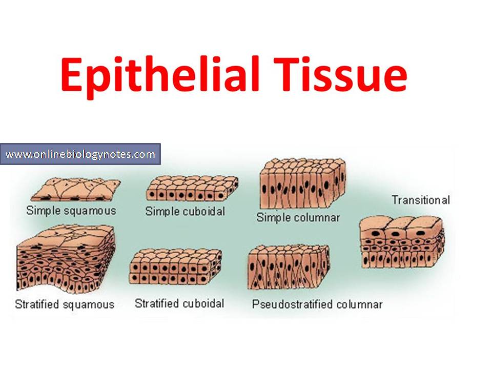 Epithelial tissue: characteristics and classification scheme and types – Online Biology NotesEpithelial tissue: characteristics and classification scheme and types epithelial tissue An epithelium is a sheet of cells that…
 My Cell.png – Google DriveMy Cell.png – Google Drive
Learn more about the effectiveness of vaccines by watching this short video!
 COVID-19 Breakthrough Cases & Vaccines – YouTubeWe’ve received many questions about breakthrough cases of COVID-19 occurring in people who have been vaccinated with one of the COVID vaccines. Dr. Matt S…
Good grades
Arteriosclerosis and Atherosclerosis
Both arteriosclerosis and atherosclerosis can cause hypertension. Arteriosclerosis is a term used to describe a disorder in which the arteries narrow and stiffen, resulting in impaired blood circulation throughout the body and increased blood pressure. Atherosclerosis is the buildup of lipid plaques,…[Read more]
[Read more]
[Read more]
[Read more]
[Read more]