-

Georgette Marie Inoferio posted an update 2 years, 7 months ago
Histology Slides (Drawings)
MT30 – FHISTO DRAWINGS – Google DriveHISTO DRAWINGS – Google Drive -

John Carlo Lumagod posted an update 2 years, 7 months ago
Histology Art | MT 30 (LAB) – F
Good day!
Attached below is my illustration of five Blood-Vascular System microscopic photographs. -
John Carlo Lumagod posted an update 2 years, 7 months ago
Histology Art | MT 30 (LAB) – F
Good day!
Attached below is my illustration of five Digestive System microscopic photographs. -
John Carlo Lumagod posted an update 2 years, 7 months ago
Histology Art | MT 30 (LAB) – F
Good day!
Attached below is my illustration of five Integumentary System microscopic photographs. -
John Carlo Lumagod posted an update 2 years, 7 months ago
Histology Art | MT 30 (LAB) – F
Good day!
Attached below is my illustration of five Nervous Tissue microscopic photographs. -
John Carlo Lumagod posted an update 2 years, 7 months ago
Histology Art | MT 30 (LAB) – F
Good day!
Attached below is my illustration of five Muscle Tissue microscopic photographs. -
John Carlo Lumagod posted an update 2 years, 7 months ago
Histology Art | MT 30 (LAB) – F
Good day!
Attached below is my illustration of five Connective Tissue microscopic photographs. -
John Carlo Lumagod changed their profile picture 2 years, 7 months ago
-
Georgette Marie Inoferio and
 Ariel Lester, Jr. Am-is are now friends 2 years, 8 months ago
 -

Jessie S. Gabay posted an update in the group
 HISTOLOGY ART – F 2 years, 8 months agoExamples of Muscle Tissue microscopic photographs.
Illustrated by: Jessie S. Gabay
Apps used: GoodNotes 5 & Canva -
Jessie S. Gabay posted an update in the group
HISTOLOGY ART – F 2 years, 9 months agoExamples of Connective Tissue microscopic photographs.
Illustrated by: Jessie S. Gabay
Apps used: GoodNotes 5 & Canva -

Misty Cadalso posted an update in the group
 HISTOLOGY ART H 2 years, 10 months agoEpithelial Tissues
 -

Kiara Formento posted an update in the group
Histology Art (MT 30 LAB C) 2 years, 10 months agoEpithelial Tissues
Illustrated by: Kiara Nathe Marie Formento -
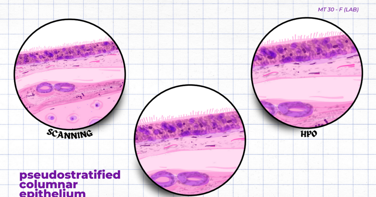
John Carlo Lumagod posted an update in the group
HISTOLOGY ART – F 2 years, 10 months agoGood day!
Attached below is my illustration of five Epithelial Tissue microscopic photographs.
Edited via: Canva
Reference:… -
John Carlo Lumagod posted an update in the group
HISTOLOGY ART – F 2 years, 10 months ago -

Reign Gacasan posted an update in the group
 Histology Art MT 30 – B 2 years, 10 months agoReign Francis A. Gacasan
MT 30 (LAB) – B
Epithelial Tissue -

Szymon Cuasito posted an update in the group
Histology Art MT 30 – B 2 years, 10 months agoSzymon Johar C. Cuasito
MT 30 LAB – B
Epithelial Tissues -

John Lloyd Fundador Baroro posted an update in the group
Histology Art MT 30 – B 2 years, 10 months agoEpithelial Tissues
John Lloyd F. Baroro
MT 30 – B
Traditional DrawingGoogle Drive: Sign-inAccess Google Drive with a Google account (for personal use) or Google Workspace account (for business use). -
Jessie S. Gabay posted an update in the group
HISTOLOGY ART – F 2 years, 10 months agoExamples of Epithelial Tissue microscopic photographs.
Illustrated by: Jessie S. Gabay
Apps used: GoodNotes 5 & CanvaGABAY_epithelial.pdf – Google… -
Misty Cadalso posted an update in the group
HISTOLOGY ART H 2 years, 10 months agoMy own interpretation of cell - Load More